ALLEN CELL EXPLORER
  • About
      Institute
      1. Our science: CellScapes
      2. Past foundational projects
      3. News feed
      4. About us
      5. Careers
  • Allen Cell Collection
      Order cells & plasmids
      1. Cell Catalog
      2. Disease Collection Cell Catalog
      3. Cell Catalog quickview
      4. Cell video shorts
      Lab methods
      1. Video protocols
      2. Written protocols
      3. Our methodology
      4. Support forum
      About our hiPS cells
      1. hiPS Cell Structure Overview
      2. Visual Guide to Human Cells
      3. Cell structure observations
      4. Why endogenous tagging?
      5. Differentiation into cardiomyocytes
      6. Genomics
      7. Download cell data: Images, genomics, & features
  • Data & Digital Tools
      General
      1. Tools & resources overview
      2. Download cell data (images, genomics, features)
      3. Code repositories & software
      Desktop tools
      1. Allen Cell & Structure Segmenter
      2. AGAVE 3D pathtrace image viewer
      Web tools
      1. BioFile Finder
      2. Cell Feature Explorer
      3. Integrated Mitotic Stem Cell
      4. └ Z-stack viewer
      5. └ 3D viewer
      Web tools (con't)
      1. Simularium viewer
      2. Timelapse Feature Explorer
      3. Visual Guide to Human Cells
      4. Vol-E (Web Volume Viewer)
      5. 3D Cell Viewer
  • Analysis & Modeling
      Allen Integrated Cell models
      1. Visual Guide to Human Cells
      2. Integrated Mitotic Stem Cell
      3. └ Z-stack viewer
      4. └ 3D viewer
      5. Allen Integrated Cell
      6. └ 3D Probabilistic Modeling
      7. └ Label-free Determination
      4D biology models
      1. Simularium viewer
      Methodologies
      1. Drug perturbation pilot study
      2. hiPS cells during mitosis
      3. Differentiation into cardiomyocytes
  • Publications
      Articles
      1. Publications
      2. Preprints
      Presentations
      1. Talks & posters
  • Education
      Educational resources
      1. All resources
      2. Teaching materials
      Online tools popular with teachers
      1. Visual Guide to Human Cells
      2. Integrated Mitotic Stem Cell
      3. 3D Cell Feature Explorer
      4. 3D Cell Viewer
      5. hiPS cell structure overview
  • Support
      Questions
      1. FAQs
      2. Forum
      Tutorials for digital tools
      1. Video tutorials
      2. Visual Guide tutorial
      3. AGAVE documentation
      Lab methods
      1. Video protocols
      2. Written protocols
      3. Our methodology
  • 🔍
      SEARCHBAR

Gene-edited cells differentiated
​into cardiomyocytes

Stem cells differentiated into cardiomyocytes beat spontaneously
 

Publication

Cell states beyond transcriptomics: integrating structural organization and gene expression in hiPSC-derived cardiomyocytes
We present a quantitative co-analysis of RNA abundance and sarcomere organization in single cells and an integrated framework to predict subcellular organization states from gene expression. This study establishes a framework for multi-dimensional analysis of single cells to study the relationships between gene expression and subcellular organization and to develop a more nuanced description of cell states.

Cell line used in the study to produce the dataset for all analyses: ACTN2-GFP (cell line ID:
AICS-0075 cl.85) - more information about the cell line can be found on the Cell Catalog page. 
BioRxiv preprint
Dataset for all analyses
Analysis code on GitHub
GitHub notebook for figures
Graphical Abstract: Transcriptional profiling and structural classification was performed on human induced pluripotent stem cell-derived cardiomyocytes to characterize the relationship between transcript abundance and subcellular organization.
Graphical Abstract

Gene edited cells differentiate into cardiomyocytes

A. 
B. 
C. 
Figure 1. After differentiation of the edited stem cells into cardiomyocytes, cells begin to beat spontaneously. Three videos taken at day 16 of differentiation show a variety of different rates and behaviors: (A) small contractile pulsing of the tissue, which remains attached to the bottom of the culture dish. (B) Wavelike, synchronous beating throughout the cardiac tissue, which has lifted off the bottom of the culture dish in most regions in this field of view. (C) Similar wavelike beating as seen in (B), however more vigorous and at a faster rate.

Playback speed is real time and each video is roughly 2.5mm across the field of view, taken from a standard camera looking through the microscope eyepiece.
Picture
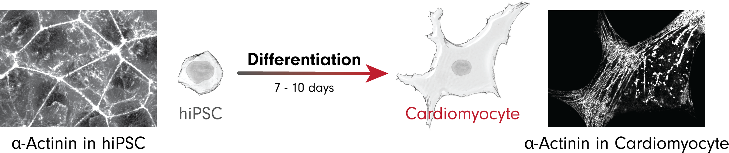
Figure 2. Many structures reorganize during differentiation. α-actinin found primarily in the rings and fiber bundles at the top and bottom of hiPSCs respectively are reorganized into the contractile apparatus of sarcomeres in cardiomyocytes.

The Institute

Home
Careers
Culture & Community
Archived Content

Legal

Terms of Use
Citation Policy
Privacy Policy
Cookie Settings

Help & contact

​FAQs
Help
​Send us a Message
​Sign up for our Newsletter
Allen Institute for Cell Science is a part of the Allen Institute. The mission of the Allen Institute is to understand the principles that govern life, and to advance health. Our creative and multi-dimensional teams focus on answering some of the biggest questions in bioscience. We accelerate foundational research, catalyze bold ideas, develop tools and models, and openly share our science to make a broad, transformational impact on the world.
Follow Us  
​Copyright © 2025 Allen Institute. All Rights Reserved.
  • About
      Institute
      1. Our science: CellScapes
      2. Past foundational projects
      3. News feed
      4. About us
      5. Careers
  • Allen Cell Collection
      Order cells & plasmids
      1. Cell Catalog
      2. Disease Collection Cell Catalog
      3. Cell Catalog quickview
      4. Cell video shorts
      Lab methods
      1. Video protocols
      2. Written protocols
      3. Our methodology
      4. Support forum
      About our hiPS cells
      1. hiPS Cell Structure Overview
      2. Visual Guide to Human Cells
      3. Cell structure observations
      4. Why endogenous tagging?
      5. Differentiation into cardiomyocytes
      6. Genomics
      7. Download cell data: Images, genomics, & features
  • Data & Digital Tools
      General
      1. Tools & resources overview
      2. Download cell data (images, genomics, features)
      3. Code repositories & software
      Desktop tools
      1. Allen Cell & Structure Segmenter
      2. AGAVE 3D pathtrace image viewer
      Web tools
      1. BioFile Finder
      2. Cell Feature Explorer
      3. Integrated Mitotic Stem Cell
      4. └ Z-stack viewer
      5. └ 3D viewer
      Web tools (con't)
      1. Simularium viewer
      2. Timelapse Feature Explorer
      3. Visual Guide to Human Cells
      4. Vol-E (Web Volume Viewer)
      5. 3D Cell Viewer
  • Analysis & Modeling
      Allen Integrated Cell models
      1. Visual Guide to Human Cells
      2. Integrated Mitotic Stem Cell
      3. └ Z-stack viewer
      4. └ 3D viewer
      5. Allen Integrated Cell
      6. └ 3D Probabilistic Modeling
      7. └ Label-free Determination
      4D biology models
      1. Simularium viewer
      Methodologies
      1. Drug perturbation pilot study
      2. hiPS cells during mitosis
      3. Differentiation into cardiomyocytes
  • Publications
      Articles
      1. Publications
      2. Preprints
      Presentations
      1. Talks & posters
  • Education
      Educational resources
      1. All resources
      2. Teaching materials
      Online tools popular with teachers
      1. Visual Guide to Human Cells
      2. Integrated Mitotic Stem Cell
      3. 3D Cell Feature Explorer
      4. 3D Cell Viewer
      5. hiPS cell structure overview
  • Support
      Questions
      1. FAQs
      2. Forum
      Tutorials for digital tools
      1. Video tutorials
      2. Visual Guide tutorial
      3. AGAVE documentation
      Lab methods
      1. Video protocols
      2. Written protocols
      3. Our methodology
  • 🔍
      SEARCHBAR