ALLEN CELL EXPLORER
  • About
      Institute
      1. News feed
      2. What we do
      3. Publications
      4. Allen Institute | allenInstitute.org
      5. Careers
      Site
      1. Home page
      2. Site updates
      3. Archived content
  • Allen Cell Collection
      Order cells & plasmids
      1. Cell Catalog
      2. Cell Catalog quickview
      3. Cell Shorts (documentaries on labs using our cells)
      4. Support forum
      Lab methods
      1. Instructional videos for success in the lab
      2. Standard operating procedures (written methods)
      3. Illustrated overviews
      About our hiPS cells
      1. hiPS Cell Structure Overview
      2. Visual Guide to Human Cells
      3. Cell structure observations
      4. Why endogenous tagging?
      5. Differentiation into cardiomyocytes
      6. Genomics
      7. Download cell data (images, genomics, features)
  • Data & Digital Tools
      Online image analysis
      1. Cell Feature Explorer (plotting & 3D viewer)
      2. 3D cell viewer (pre2018)
      3. Deep cell zoom (216,016 cells)
      Online modeling viewers
      1. Visual Guide to Human Cells
      2. Simularium (4D visual analysis)
      3. Integrated Mitotic Stem Cell
      4. └ Z-stack viewer
      5. └ 3D viewer
      6. Allen Integrated Cell viewer
      7. Label-free examples viewer
      8. 3D probabilistic model viewer
      Desktop tools
      1. Allen Cell & Structure Segmenter
      2. AGAVE 3D pathtrace image viewer
      Data & code
      1. Download cell data (images, genomics, features)
      2. Code repositories & software
  • Analysis & Modeling
      Allen Integrated Cell models
      1. Overview
      2. Integrated Mitotic Stem Cell
      3. └ Z-stack viewer
      4. └ 3D viewer
      5. Label-free Determination
      6. └ 3D viewer
      7. 3D Probabilistic Modeling
      8. └ 3D viewer
      9. Visual Guide to Human Cells
      4D biology models
      1. Simularium (online 4D viewer)
      Methodologies
      1. Drug perturbation pilot study
      2. hiPS cells during mitosis
      3. Differentiation into cardiomyocytes
  • Publications
      Articles
      1. All journal publications
      2. Preprints (biorxiv, arxiv)
      Posters
      1. Select posters
  • Education
      Education resources
      1. All Resources
      2. Teaching materials
      Online tools popular with teachers
      1. Visual Guide to Human Cells
      2. Integrated Mitotic Stem Cell
      3. Cell Feature Explorer (interactive plotting & 3D viewer)
      4. 3D cell viewer (pre2018 data)
      5. hiPS cell structure overview
  • Support
      Questions
      1. FAQs
      2. Forum
      Tutorials for digital tools
      1. Digital tool tutorials with videos
      2. Visual Guide tutorial
      3. AGAVE user guide
      Lab methods
      1. Instructional videos for success in the lab
      2. Standard operating procedures (written methods)
      3. Illustrated overviews
  • 🔍
      SEARCHBAR

Label-free Determination

​Image with unprecedented clarity compared to other tagging methods​
Press Announcement
Nature Method article
GitHub
Data Download

Label-free determination of human stem cell organization

As part of the Allen Integrated Cell, we have developed and implemented a label-free prediction method for determining the location of 3D structures inside the cell, directly from transmitted light images.
Picture
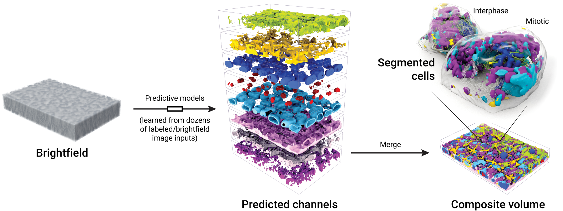
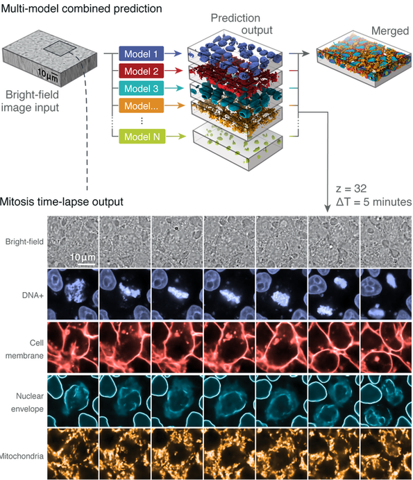
Figure 1. Images from our collection of cells can be used to generate models that produce integrated images of 3D subcellular structures—all from an input brightfield image of a field of cells. Cell segmentations can then be applied to the predictions to determine structure localization for individual cells.
Various imaging methods are currently used to capture details of subcellular organization. These, however, present trade-offs:
  • fluorescence microscopy allows the visualization of specific structures, but is both expensive and time consuming
  • live cells can be damaged by microscope laser light
  • the number of fluorescent labels which can be used at the same time is restricted by the current technology
  • the quantity of tagged structures which may be simultaneously imaged is limited​
Therefore, it is difficult to achieve an integrated representation of cellular organization using current methods. The Label-Free Determination model can leverage the specificity of fluorescence microscopy while removing many restrictions on numbers of simultaneous labels, and presents a potentially important tool for biologists for achieving insight into the integrated activities of subcellular structures.
Nature Method article
GitHub
Explore how several organelles interact in the human induced pluripotent stem cell in several individual real cells in the viewer below:

Label-free determination 3D cell structure viewer

Picture
Open Viewer

How does the label-free determination model work?

The “discrete” label-free structure determination tool consists of a convolutional neural network (CNN)-based method (see Label-free imaging tool pipeline section below), employing a U-Net architecture to model the relationships between 3D transmitted light (brightfield) and fluorescence images corresponding to several major subcellular structures (e.g. nuclear envelope, nucleoli, endoplasmic reticulum, mitochondria, etc) tagged with fluorescent proteins (see What we do, cell methods, and microscopy methods pages for further detail). The tool can train a model to learn this relationship for the structure of interest given only spatially registered pairs of images, even with a relatively small image set for training (30 image pairs per structure). The resultant model can, in turn, be used to predict a 3D fluorescence image from a new transmitted light input.

​Model predictions for a variety of subcellular structures can be combined, enabling multi-channel, integrated fluorescence imaging from a single transmitted light input.

Figure 2. ​Label-free determination imaging pipeline

Picture
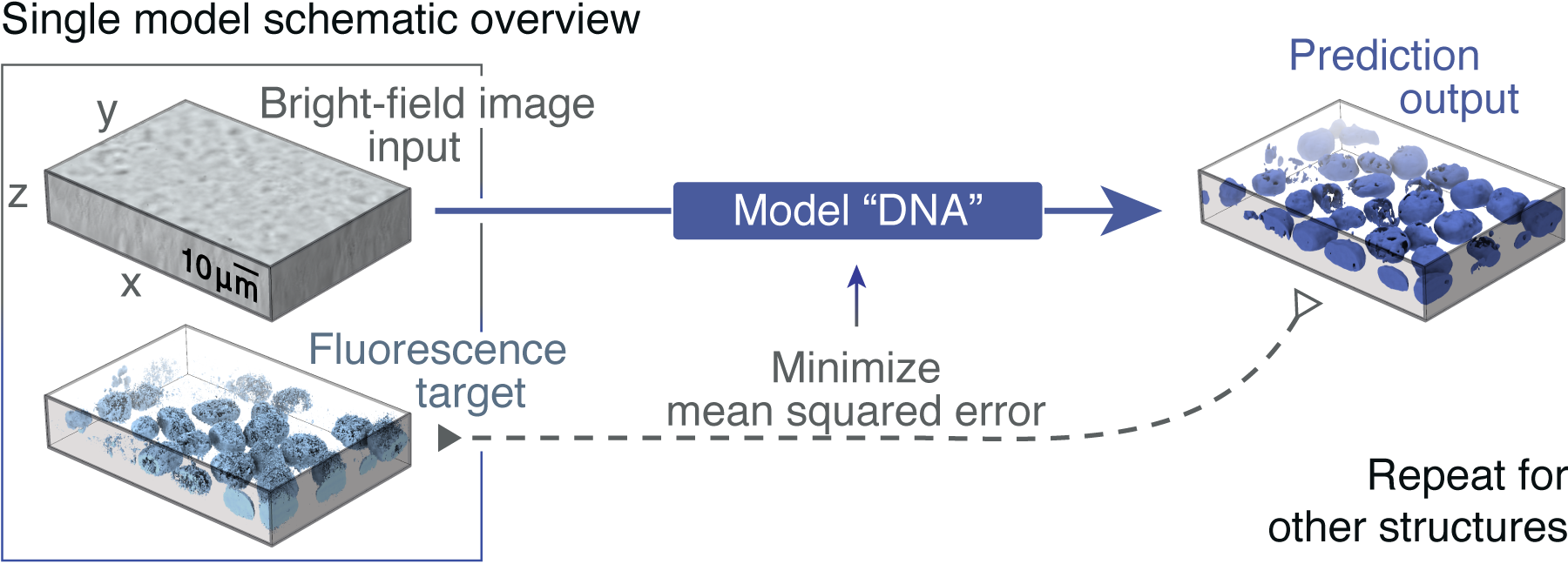
Figure 2a. Given the input of a transmitted light and fluorescence image pairs, the model is trained to minimize the mean squared error (MSE) between the fluorescence ground-truth and output of the model.

Picture
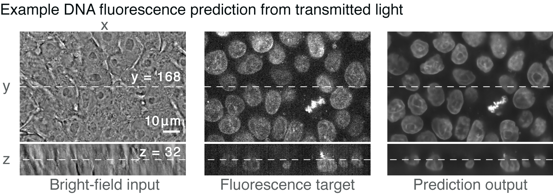
Figure 2b. Example of a 3D input transmitted light image, a ground-truth confocal DNA fluorescence image, and a tool prediction.

Picture
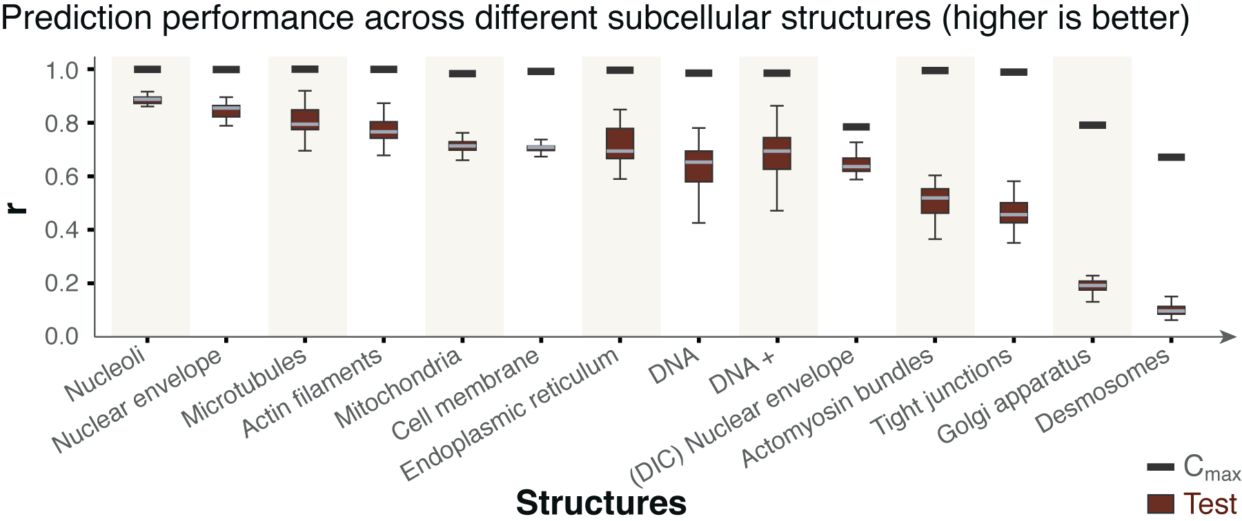
Figure 2c. Distributions of the image-wise correlation coefficient (r) between target and predicted test images from models trained on 30 3D images for the indicated subcellular structure, plotted as a box across 25th, 50th and 75th percentile, with whiskers indicating the last data points within 1.5 interquartile range of the lower and upper quartiles. For a complete description of structure labels, see Publications. ​​Black bars indicate maximum correlation between the target fluorescence image and a theoretical, noise-free image (Cmax; details of metric in Publications). 

Picture
Figure 2d. Different models applied to the same input and combined to predict multiple imaging modes. Predicted localization of DNA (blue), cell membrane (red), nuclear envelope (cyan) and mitochondria (orange) of a sample taken at 5-minute intervals. The center z-slice is shown. A mitotic event, along with stereotypical reorganization of subcellular structures, is clearly observed. 

Label-free determination over time

Figure 3. ​Notably, models trained on static images (from a single time point) can be used to predict 3D fluorescence time-lapse movies, given a 3D transmitted light input (Figure 2 panel e). See Modeling Publications for additional information.

Label-free Data Download

Label-free Prediction Training Data
​The images used to train models described above are provided here.
Label-free Prediction Training Data Download

Label-Free Imaging Collection
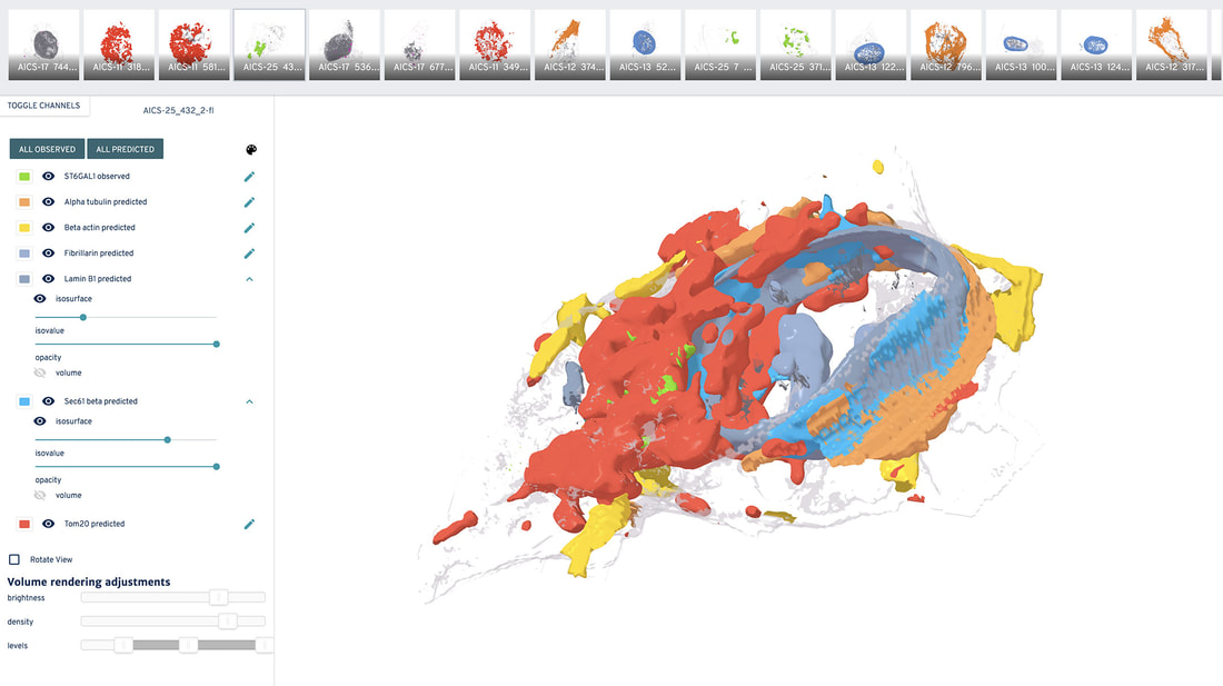
​The label-free method is applied to our entire publicly available collection of images, and the images are provided here.

​Note:
This data is intended for scientific exploration and the accuracy and utility of the predictions must be assessed structure by structure in the context of the intended use case.
Label-free imaging collection data Download

Other resources

Modeling Publications
The latest publications & preprints related to the 3D Statistical Cell Model
Methods for Modeling
Detailed and technical methods from the publications provided here
Software & Code
Links to our repositories, where all our open-source tools and code can be found.

Home

Terms of Use

Citation Policy

Privacy Policy

FAQs

Help

Send us a message

Our diversity, equity & inclusion commitment
Copyright © 2023 Allen Institute. All Rights Reserved.
cellscience.alleninstitute.org
  • About
      Institute
      1. News feed
      2. What we do
      3. Publications
      4. Allen Institute | allenInstitute.org
      5. Careers
      Site
      1. Home page
      2. Site updates
      3. Archived content
  • Allen Cell Collection
      Order cells & plasmids
      1. Cell Catalog
      2. Cell Catalog quickview
      3. Cell Shorts (documentaries on labs using our cells)
      4. Support forum
      Lab methods
      1. Instructional videos for success in the lab
      2. Standard operating procedures (written methods)
      3. Illustrated overviews
      About our hiPS cells
      1. hiPS Cell Structure Overview
      2. Visual Guide to Human Cells
      3. Cell structure observations
      4. Why endogenous tagging?
      5. Differentiation into cardiomyocytes
      6. Genomics
      7. Download cell data (images, genomics, features)
  • Data & Digital Tools
      Online image analysis
      1. Cell Feature Explorer (plotting & 3D viewer)
      2. 3D cell viewer (pre2018)
      3. Deep cell zoom (216,016 cells)
      Online modeling viewers
      1. Visual Guide to Human Cells
      2. Simularium (4D visual analysis)
      3. Integrated Mitotic Stem Cell
      4. └ Z-stack viewer
      5. └ 3D viewer
      6. Allen Integrated Cell viewer
      7. Label-free examples viewer
      8. 3D probabilistic model viewer
      Desktop tools
      1. Allen Cell & Structure Segmenter
      2. AGAVE 3D pathtrace image viewer
      Data & code
      1. Download cell data (images, genomics, features)
      2. Code repositories & software
  • Analysis & Modeling
      Allen Integrated Cell models
      1. Overview
      2. Integrated Mitotic Stem Cell
      3. └ Z-stack viewer
      4. └ 3D viewer
      5. Label-free Determination
      6. └ 3D viewer
      7. 3D Probabilistic Modeling
      8. └ 3D viewer
      9. Visual Guide to Human Cells
      4D biology models
      1. Simularium (online 4D viewer)
      Methodologies
      1. Drug perturbation pilot study
      2. hiPS cells during mitosis
      3. Differentiation into cardiomyocytes
  • Publications
      Articles
      1. All journal publications
      2. Preprints (biorxiv, arxiv)
      Posters
      1. Select posters
  • Education
      Education resources
      1. All Resources
      2. Teaching materials
      Online tools popular with teachers
      1. Visual Guide to Human Cells
      2. Integrated Mitotic Stem Cell
      3. Cell Feature Explorer (interactive plotting & 3D viewer)
      4. 3D cell viewer (pre2018 data)
      5. hiPS cell structure overview
  • Support
      Questions
      1. FAQs
      2. Forum
      Tutorials for digital tools
      1. Digital tool tutorials with videos
      2. Visual Guide tutorial
      3. AGAVE user guide
      Lab methods
      1. Instructional videos for success in the lab
      2. Standard operating procedures (written methods)
      3. Illustrated overviews
  • 🔍
      SEARCHBAR