Publications
A comprehensive list of publications collected from each menu category

|
Establishing a conceptual framework for holistic cell states and state transitions
Rafelski SM, Theriot JA. Cell, May 2024. |

|
Incomplete-penetrant hypertrophic cardiomyopathy MYH7 G256E mutation causes hypercontractility and elevated mitochondrial respiration
Lee S, Vander Roest AS, Blair CA, Kao K, Bremner SB, Childers MC, Pathak D, Heinrich P, Lee D, Chirikian O, Mohran SE, Roberts B, Smith JE, Jahng JW, Paik DT, Wu JC, Gunawardane RN, Ruppel KM, Mack DL, Pruitt BL, Regnier M, Wu SM, Spudich JA, Bernstein D. PNAS, April 2024. |

|
Understanding metric-related pitfalls in image analysis validation
Reinke A, Tizabi MD, Baumgartner M, Eisenmann M, Heckmann-Nötzel D, Kavur AE, Rädsch T, Sudre CH, Acion L, Antonelli M, Arbel T, Bakas S, Benis A, Buettner F, Cardoso MJ, Cheplygina V, Chen J, Christodoulou E, Cimini BA, Farahani K, Ferrer L, Galdran A, van Ginneken B, Glocker B, Godau P, Hashimoto DA, Hoffman MM, Huisman M, Isensee F, Jannin P, Kahn CE, Kainmueller D, Kainz B, Karargyris A, Kleesiek J, Kofler F, Kooi T, Kopp-Schneider A, Kozubek, M, Kreshuk A, Kurc T, Landman BA, Litjens G, Madani A, Maier-Hein K, Martel AL, Meijering E, Menze B, Moons KGM, Müller H, Nichyporuk B, Nickel F, Petersen J, Rafelski SM, Rajpoot N, Reyes M, Riegler MA, Rieke N, Saez-Rodriguez J, Sánchez CI, Shetty S, Summers RM, Taha AA, Tiulpin A, Tsaftaris SA, Van Calster B, Varoquaux G, Yaniv ZR, Jäger PF, Maier-Hein L. Nature Methods, February 2024. |

|
Community-developed checklists for publishing images and image analyses
Schmied C, Nelson MS, Avilov S, Bakker GJ, Bertocchi C, Bischof J, Boehm U, Brocher J, Carvalho MT, Chiritescu C, Christopher J, Cimini BA, Conde-Sousa E, Ebner M, Ecker R, Eliceiri K, Fernandez-Rodriguez J, Gaudreault N, Gelman L, Grunwald D, Gu T, Halidi N, Hammer M, Hartley M, Held M, Jug F, Kapoor V, Koksoy AA, Lacoste J, Dévédec SL, Guyader SL, Liu P, Martins GG, Mathur A, Miura K, Llopis PM, Nitschke R, North A, Parslow AC, Payne-Dwyer A, Plantard L, Ali R, Schroth-Diez B, Schütz L, Scott RT, Seitz A, Selchow O, Sharma VP, Spitaler M, Srinivasan S, Strambio-De-Castillia C, Taatjes D, Tischer C, Jambor HK. Nature Methods, February 2024. |

|
Automated human inducible pluripotent stem cell culture and sample preparation for 3D live cell microscopy
Gregor BW, Coston ME, Adams EM, Arakaki J, Borensztejn A, Do TP, Fuqua MA, Haupt A, Hendershott MC, Leung W, Mueller IA, Nath A, Nelson AM, Rafelski SM, Sanchez EE, Swain-Bowden MJ, Tang WJ, Thirstrup DJ, Wiegraebe W, Whitney BP, Yan C, Gunawardane RN, Gaudreault N. Nature Protocols, December 2023. |

|
Building the next generation of virtual cells to understand cellular biology
Johnson GT, Agmon E, Akamatsu M, Lundberg E, Lyons B, Ouyang W, Quintero-Carmona OA, Riel-Mehan M, Rafelski SM, Horwitz R. Biophysical Journal, September 19, 2023. |
|
OME-Zarr: a cloud-optimized bioimaging file format with international community support
Moore J, Basurto-Lozada D, Besson S, Bogovic J, Bragantini J, Brown EM, Burel JM, Monero XC, Medeiros G, Diel EE, Gault D, Ghosh SS, Gold I, Halchenko YO, Hartley M, Horsfall D, Keller MS, Kittisopikul MS, Kovacs G, Yoldaş AK, Kyoda K, Villegeorges AT, Li T, Liberali P, Linkert M, Lindner D, Linkert M, Lüthi J, Maitin-Shepard J, Manz T, Marconato L, McCormick M, Lange M, Mohamed K, Moore W, Norlin N, Ouyang W, Özdemir B, Palla G, Pape C, Pelkmans L, Pietzsch T, Preibisch S, Prete M, Rzepka N, Samee S, Schaub N, Sidky H, Solak AC, Stirling DR, Striebel J, Tischer C, Toloudis D, Virshup I, Walczysko P, Watson AM, Weisbart E, Wong F, Yamauchi KA, Bayraktar O, Cimini BA, Gehlenborg N, Haniffa M, Hotaling N, Onami S, Royer LA, Saalfeld S, Stegle O, Theis FJ, & Swedlow JR. Histochemistry and Cell Biology, July 10, 2023. |

|
Spatial and temporal organization of the genome: Current state and future aims of the 4D nucleome project
Dekker J, Alber F, Aufmkolk S, Beliveau BJ, Bruneau BG, Belmont AS, Bintu L, Boettiger A, Calandrelli R, Disteche CM, Gilbert DM, Gregor T, Hansen AS, Huang B, Huangfu D, Kalhor R, Leslie CS, Li W, Li Y, Ma J, Noble WS, Park PJ, Phillips-Cremins JE, Pollard KS, Rafelski SM, Ren B, Ruan Y, Shav-Tal Y, Shen Y, Shendure J, Shu X, Strambio-De-Castillia C, Vertii A, Zhang H, Zhong S. Molecular Cell, July 6, 2023. |

|
When seeing is not believing: application-appropriate validation matters for quantitative bioimage analysis
Chen J, Viana MP, Rafelski SM. Nature Methods, July 11, 2023. |

|
Reporting reproducible imaging protocols
Larsen DD, Gaudreault N, Gibbs HC. STAR Protocols, March 2, 2023. |

|
Integrated intracellular organization and its variations in human iPS cells
Viana MP, Chen J, Knijnenburg TA, Vasan R, Yan C, Arakaki JE, Bailey M, Berry B, Borensztejn A, Brown EM, Carlson S, Cass JA, Chaudhuri B, Metzler KRC, Coston ME, Crabtree ZJ, Davidson S, DeLizo CM, Dhaka S, Dinh SQ, Do TP, Domingus J, Donovan-Maiye RM, Ferrante AJ, Foster TJ, Frick CL, Fujioka G, Fuqua MA, Gehring JL, Gerbin KA, Grancharova T, Gregor BW, Harrylock L, Haupt A, Hendershott MC, Hookway C, Horwitz AR, Hughes C, Isaac EJ, Johnson GR, Kim B, Leonard AN, Leung W, Lucas JJ, Ludmann SA, Lyons BM, Malik H, McGregor R, Medrash GE, Meharry SL, Mitcham K, Mueller IA, Murphy-Stevens TL, Nath A, Nelson AM, Oluoch SA, Paleologu L, Popiel TA, Riel-Mehan MM, Roberts B, Schaefbauer LM, Schwarzl M, Sherman J, Slaton S, Sluzewski MF, Smith JE, Sul Y, Swain-Bowden MJ, Tang WJ, Thirstrup DJ, Toloudis DT, Tucker AP, Valencia V, Wiegraebe W, Wijeratna T, Yang R, Zaunbrecher RJ, Labitigan RLD, Sanborn AL, Johnson GT, Gunawardane RN, Gaudreault N, Theriot JA, Rafelski SM. Nature, January 4, 2023. |

|
The dawn of interoperating spatial models in cell biology
Iwasa JH, Lyons B, Johnson GT. Elsevier, December 2022. |
|
The new era of quantitative cell imaging—challenges and opportunities
Bagheri N, Carpenter AE , Lundberg E, Plant AL , Horwitz R. Molecular Cell, January 20, 2022. |
|
The Simularium Viewer: an interactive online tool for sharing spatiotemporal biological models
Lyons B, Isaac E, Choi NH, Do TP, Domingus J, Iwasa J, Leonard A, Riel-Mehan M, Rodgers E, Schaefbauer L, Toloudis D, Waltner O, Wilhelm L, Johnson GT. Nature Methods, April 04, 2022. |
|
A deep generative model of 3D single-cell organization
Donovan-Maiye RM, Brown JM, Chan CK, Ding L, Yan C, Gaudreault N, Theriot JA, Maleckar MM, Knijnenburg TA, Johnson GR. PLOS Computational Biology, January 18, 2022. |
|
|
Does Organelle Shape Matter?: Exploring Patterns in Cell Shape and Structure with High-Throughput (HT) Imaging
Goller CC, Johnson GT, Casimo K. CourseSource, 2022. |
|
It’s time to incorporate diversity into our basic science and disease models
Horwitz R, Riley EA, Millan MT, Gunawardane RN. Nature Cell Biology, November 29, 2021. |
|
QUAREP-LiMi: A community-driven initiative to establish guidelines for quality assessment and reproducibility for instruments and images in light microscopy
Nelson G, Boehm U, Bagley S, Bajcsy P, Bischof J, Brown CM, Dauphin A, Dobbie IM, Eriksson JE, Faklaris O, Fernandez-Rodriguez J, Ferrand A, Gelman L, Gheisari A, Hartmann H, Kukat C, Laude A, Mitkovski M, Munck S, North AJ, Rasse TM, Resch-Genger U, Schuetz LC, Seitz A, Strambio-De-Castillia C, Swedlow JR, Alexopoulos I, Aumayr K, Avilov S, Bakker GJ, Bammann RR, Bassi A, Beckert H, Beer S, Belyaev Y, Bierwagen J, Birngruber KA, Bosch M, Breitlow J, Cameron LA, Chalfoun J, Chambers JJ, Chen CL, Conde-Sousa E, Corbett AD, Cordelieres FB, Nery ED, Dietzel R, Eismann F, Fazeli E, Felscher A, Fried H, Gaudreault N, Goh WI, Guilbert T, Hadleigh R, Hemmerich P, Holst GA, Itano MS, Jaffe CB, Jambor HK, Jarvis SC, Keppler A, Kirchenbuechler D, Kirchner M, Kobayashi N, Krens G, Kunis S, Lacoste J, Marcello M, Martins GG, Metcalf DJ, Mitchell CA, Moore J, Mueller T, Nelson MS, Ogg S, Onami S, Palmer AL, Paul-Gilloteaux P, Pimentel JA, Plantard L, Podder S, Rexhepaj E, Royon A, Saari MA, Schapman D, Schoonderwoert V, Schroth-Diez B, Schwartz S, Shaw M, Spitaler M, Stoeckl MT, Sudar D, Teillon J, Terjung S, Thuenauer R, Wilms CD, Wright GD, Nitschke R. Journal of Microscopy, Volume 284, Issue 1, July 02, 2021. |
|
A comprehensive analysis of gene expression changes in a high replicate and open-source dataset of differentiating hiPSC-derived cardiomyocytes
Grancharova T, Gerbin KA, Rosenberg AB, Roco CM, Arakaki JE, DeLizo CM, Dinh SQ, Donovan-Maiye RM, Hirano M, Nelson AM, Tang J, Theriot JA, Yan C, Menon V, Palecek SP, Seelig G, Gunawardane RN. Scientific Reports, August 04, 2021. |
|
Cell states beyond transcriptomics: Integrating structural organization and gene expression in hiPSC-derived cardiomyocytes
Gerbin KA, Grancharova T, Donovan-Maiye RM, Hendershott MC, Anderson HG, Brown JM, Chen J, Dinh SQ, Gehring JL, Johnson GR, , Lee H, Nath A, Nelson AM, Sluzewski MF, Viana MP, Yan C, Zaunbrecher RJ, Cordes Metzler KR, Gaudreault N, Knijnenburg T, Rafelski SM, Theriot JA, Gunawardane RN. Cell Systems, June 16, 2021. |

|
A Bayesian framework for the detection of diffusive heterogeneity
Cass JA, Williams CD, Theriot J. PLoS One, May 7, 2020. |

|
"Not just a cog": a Q&A on team science in cell biology
Tompa R, Rafelski S, Johnson GT.ASCB Science News, May 7, 2020. |
|
DLITE Uses Cell-Cell Interface Movement to Better Infer Cell-Cell Tensions
Vasan R, Maleckar MM, Williams CD, Rangamani P. Biophysical Journal, November 5, 2019. |

|
A virtual laboratory on cell division using a publicly-available image database
Shelden EA, Offerdahl EG, Johnson GT. CourseSource: Evidence-based teaching resources for undergraduate biology education, May 2019. |

|
Fluorescent Gene Tagging of Transcriptionally Silent Genes in hiPSCs
Roberts B, Hendershott MC, Arakaki J, Gerbin KA, Malik H, Nelson A, Gehring J, Hookway C, Ludmann SA, Yang R, Haupt A, Grancharova T, Valencia V, Fuqua MA, Tucker A, Rafelski SM, Gunawardane RN. Stem Cell Reports, May 2019. |

|
Seeing beyond sight: new computational approaches to understanding cells
Johnson GT, Rafelski S. ASCB Science News, December 20, 2018. |

|

|
Label-free prediction of three-dimensional fluorescence images from transmitted-light microscopy
Ounkomol C, Seshamani S, Maleckar MM, Collman F, Johnson GR. Nature Methods, September 7, 2018. |

|
Endogenous Protein Tagging in Human Induced Pluripotent Stem Cells Using CRISPR/Cas9
Haupt A, Grancharova T, Arakaki J, Fuqua MA, Roberts B, Gunawardane RN. Jove, August 25, 2018. |

|
CellProfiler 3.0: Next-generation image processing for biology
McQuin C, Goodman A, Chernyshev V, Kamentsky L, Cimini BA, Karhohs KW, Doan M, Ding L, Rafelski SM, Thirstrup D, Wiegraebe W, Singh S, Becker T, Caicedo JC, Carpenter AE. PLOS | Biology, July 3, 2018. |
|
Systematic gene tagging using CRISPR/Cas9 in human stem cells to illuminate cell organization
Roberts B, Haupt A, Tucker A, Grancharova T, Arakaki J, Fuqua MA, Nelson A, Hookway C, Ludmann SA, Mueller IA, Yang R, Horwitz AR, Rafelski SM, Gunawardane RN. Molecular Biology of the Cell (MBoC), August 16, 2017. |

|
Whole cell maps chart a course for 21st-century cell biology
Horwitz R, Johnson GT. Science, May 26, 2017. |

|

|

|

|
A Integrated, multi-scale, spatial–temporal cell biology – A next step in the post genomic era
Horwitz R. Methods, March 2016. |

|
Colony context and size-dependent compensation mechanisms give rise to variations in nuclear growth trajectories
Dixon JC, Frick CL, Leveille CL, Garrison P, Lee PA, Mogre SS, Morris B, Nivedita N, Vasan R, Chen J, Fraser CL, Gamlin CR, Harris LK, Hendershott MC, Johnson GT, Klein KN, Oluoch SA, Thirstrup DJ, Sluzewski MF, Wilhelm L, Yang R, Toloudis DM, Viana MP, Theriot JA, Rafelski SM. BioRXiv, June 28, 2024. |

|
Harmonizing the Generation and Pre-publication Stewardship of FAIR Image Data
Bialy N, Alber F, Andrews B, Angelo M, Beliveau B, Bintu L, Boettiger A, Boehm U, Brown CM, Maina MB, Chambers JJ, Cimini BA, Eliceiri K, Errington R, Faklaris O, Gaudreault N, Germain RN, Goscinski W, Grunwald D, Halter M, Hanein D, Hickey JW, Lacoste J, Laude A, Lundberg E, Ma J, Malacrida L, Moore J, Nelson G, Neumann EK, Nitschke R, SOnami, Pimentel JA, Plant AL, Radtke AJ, Sabata B, Schapiro D, Schöneberg J, Spraggins JM, Sudar D, Vierdag WMAM, Volkmann N, Wählby C, Wang S, Yaniv Z, Strambio-De-Castillia C. arRXiv, February 8, 2024. |

|
Enabling Global Image Data Sharing in the Life Sciences
Bajcsy P, Bhattiprolu S, Boerner K, Cimini BA, Collinson L, Ellenberg J, Fiolka R, Giger M, Goscinski W, Hartley M, Hotaling N, Horwitz R, Jug F, Kreshuk A, Lundberg E, Mathur A, Narayan K, S Onami, Plant AL, Prior F, J Swedlow, Taylor A, Keppler A. arRXiv, February 2, 2024. |

|
OME-Zarr: a cloud-optimized bioimaging file format with international community support
Moore J, Basurto-Lozada D, Besson S, Bogovic J, Brown EM, Burel JM, Medeiros G, Diel EE, Gault D, Ghosh SS, Gold I, Halchenko YO, Hartley M, Horsfall D, Keller MS, Kittisopikul MS, Kovacs G, Yoldaş AK, Villegeorges AT, Li T, Liberali P, Linkert M, Lindner D, Lüthi J, Maitin-Shepard J, Manz T, McCormick M, Mohamed K, Moore W, Özdemir B, Pape C, Pelkmans L, Prete M, Pietzsch T, Preibisch S, Rzepka N, Stirling DR, Striebel J, Tischer C, Toloudis D, Walczysko P, Watson AM, Wong F, Yamauchi KA, Bayraktar O, Haniffa M, Saalfeld S, Swedlow JR. BioRXiv, February 21, 2023. |

|
A perspective on Microscopy Metadata: data provenance and quality control
Huisman M, Hammer M, Rigano A, Boehm U, Chambers JJ, Gaudreault N, North AJ, Pimentel JA, Sudar D, Bajcsy P, Brown CM, Corbett AD, Faklaris O, Lacoste J, Laude A, Nelson G, Nitschke R, Grunwald D, Strambio-De-Castillia C. arXiv, April 26, 2021. |

|
Towards community-driven metadata standards for light microscopy: tiered specifications extending the OME model
Hammer M, Huisman M, Rigano A, Boehm U, Chambers JJ, Gaudreault N, North AJ, Pimentel JA, Sudar D, Bajcsy P, Brown CM, Corbett AD, Faklaris O, Lacoste J, Laude A, Nelson G, Nitschke R, Farzam F, Smith C, Grunwald D, Strambio-De-Castillia C. BioRXiv, April 26, 2021. |

|
Automated hiPSC culture and sample preparation for 3D live cell microscopy
Coston ME, Gregor BW, Arakaki J, Borensztejn A, Do TP, Fuqua MA, Haupt A, Hendershott MC, Leung W, Mueller IA, Nelson AM, Rafelski SM, Swain-Bowden MJ, Tang WJ, Thirstrup DJ, Wiegraebe W, Yan C, Gunawardane RN, Gaudreault N. BioRXiv, December 19, 2020. |

|
Robust integrated intracellular organization of the human iPS cell: where, how much, and how variable
Viana MP, Chen J, Knijnenburg TA, Vasan R, Yan C, Arakaki JE, Bailey M, Berry B, Borensztejn A, Brown JM, Carlson S, Cass JA, Chaudhuri B, Metzler KRC, Coston ME, Crabtree ZJ, Davidson S, DeLizo CM, Dhaka S, Dinh SQ, Do TP, Domingus J, Donovan-Maiye RM, Foster TJ, Frick CL, Fujioka G, Fuqua MA, Gehring JL, Gerbin KA, Grancharova T, Gregor BW, Harrylock L, Haupt A, Hendershott MC, Hookway C, Horwitz AR, Hughes C, Isaac EJ, Johnson GR, Kim B, Leonard AN, Leung W, Lucas JJ, Ludmann SA, Lyons BM, Malik H, McGregor R, Medrash GE, Meharry SL, Mitcham K, Mueller IA, Murphy-Stevens TL, Nath A, Nelson AM, Paleologu L, Popiel TA, Riel-Mehan MM, Roberts B, Schaefbauer LM, Schwarzl M, Sherman J, Slaton S, Sluzewski MF, Smith JE, Sul Y, Swain-Bowden MJ, Tang WJ, Thirstrup DJ, Toloudis DT, Tucker AP, Valencia V, Wiegraebe W, Wijeratna T, Yang R, Zaunbrecher RJ, Science AI for C, Johnson GT, Gunawardane RN, Gaudreault N, Theriot JA, Rafelski SM. BioRXiv, December 10, 2020. |

|
Cell states beyond transcriptomics: integrating structural organization and gene expression in hiPSC-derived cardiomyocytes
Gerbin KA, Grancharova T, Donovan-Maiye R, Hendershott MC, Brown J, Dinh SQ, Gehring JL, Hirano M, Johnson GR, Nath A, Nelson A, Roco CM, Rosenberg AB, Sluzewski MF, Viana MP, Yan C, Zaunbrecher RJ, Cordes Metzler KR, Menon V, Palecek SP, Seelig G, Gaudreault N, Knijnenburg T, Rafelski SM, Theriot JA, Gunawardane RN. BioRXiv, May 27, 2020. |

|
The Allen Cell Structure Segmenter: a new open source toolkit for segmenting 3D intracellular structures in fluorescence microscopy images
Chen J, Ding L, Viana MP, Hendershott MC, Yang R, Mueller IA, Rafelski S. BioRxiv, December 8, 2018 . |

|
Scarless gene tagging of transcriptionally silent genes in hiPSCs to visualize cardiomyocyte sarcomeres in live cells
Roberts B, Arakaki J, Gerbin KA, Malik H, Nelson A, Hendershott MC, Hookway C, Ludmann SA, Mueller IA, Yang R, Rafelski SM, Gunawardane RN. BioRxiv, June 2018. |

|
Label-free prediction of three-dimensional fluorescence images from transmitted light microscopy
Ounkomol C, Seshamani S, Maleckar MM, Collman F, Johnson GR. BioRxiv, May 2018. |

|

|
Three dimensional cross-modal image inference: label-free methods for subcellular structure prediction
Ounkomol C, Fernandes DA, Seshamani S, Maleckar MM, Collman F, Johnson GR. BioRxiv, November 2017. |

|
Final article published in Molecular Biology of the Cell (MBoC) available here.
Preprint: Systematic Gene Tagging using CRISPR/Cas9 in Human Stem Cells to Illuminate Cell Organization Roberts B, Haupt A, Tucker A, Grancharova T, Arakaki J, Fuqua MA, Nelson A, Hookway C, Ludmann SA, Mueller IA, Yang R, Horwitz AR, Rafelski SM, Gunawardane RN. BioRxiv, April 2017 |

|
Preprint: Generative Modeling with Conditional Autoencoders: Building an Integrated Cell
Johnson GR, Donavan-Maiye RM, Maleckar MM. arXiv, April 2017. |
|
|
Illumination Power and illumination stability
Gaudreault N, Bagley S, Bammann RR, Barachati F, Barna L, Boczonadi V, Boehm U, Bosch M, Brideau C, Carvalho MT, Colarusso P, Cole R, Darwish-Miranda N, Duwé S, Eismann F, Faklaris O, Felscher A, Gonnert M, Grunwald D, Kirchner M, Hoffmann B, Krens G, Laude AJ, LeDue JM, Lorentz P, Mitkovski M, Nelson MS, Schroth-Diez B, Schwartz S, Srinivasan S, Thuenauer R, Wee TL, Oord KVD, Oostende-Triplet CV, Nitschke R, Gelman L. Protocols.io, February 2022. |
|
|
Maintenance of Undifferentiated hiPSC Cultures and Differentiation to Cardiomyocytes on Glass Surfaces
Nelson A, Dinh SQ, Gerbin K, Hendershott M, Hookway C, Gunawardane RN. Protocols.io, March 2021. |
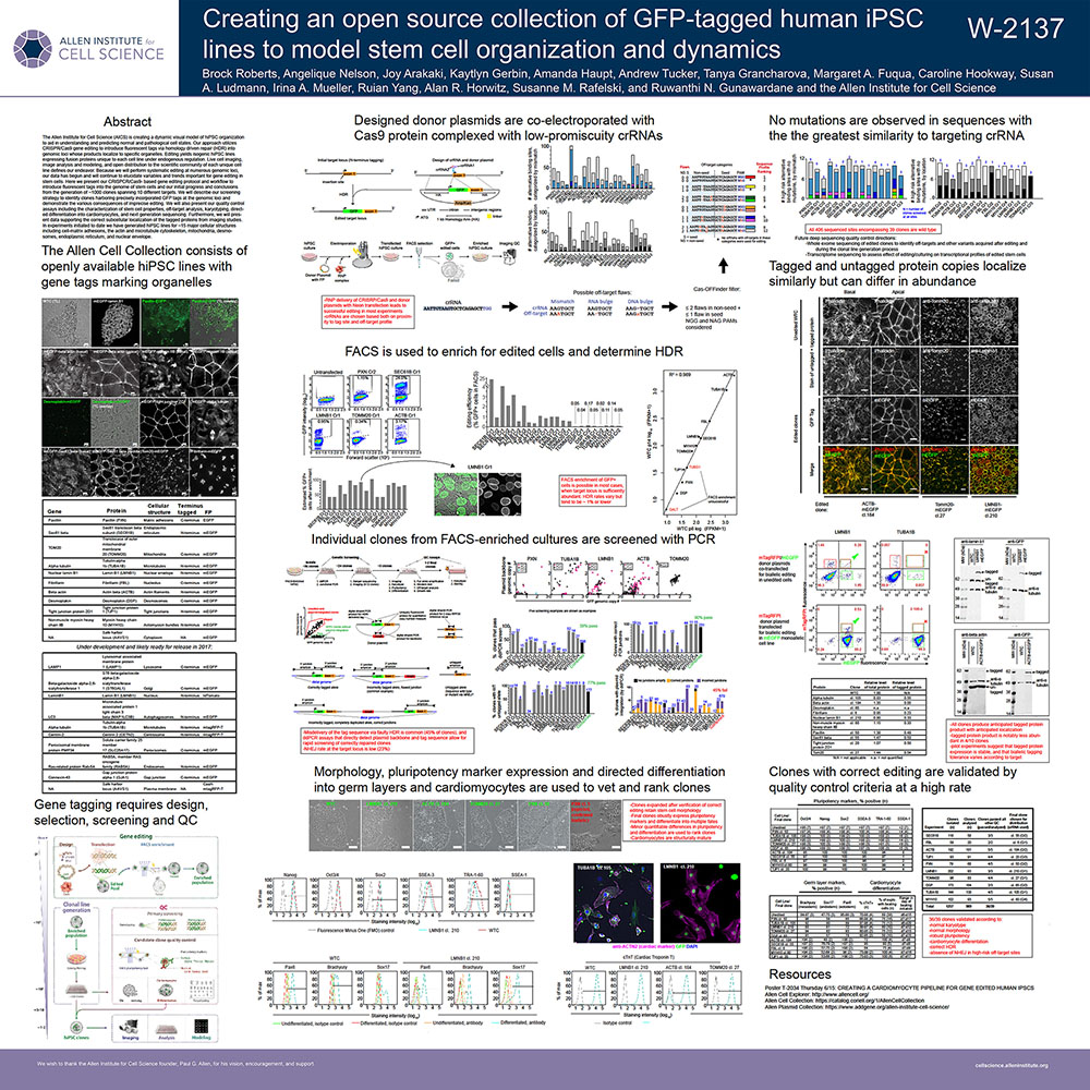
| poster-isscr_tagged-ipsc-open-source-collection_1.pdf |
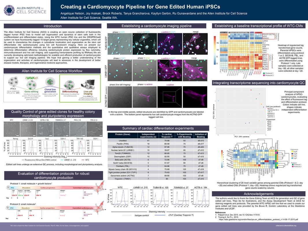
| poster-isscr_creating-a-cardiomyocyte-pipeline-for_gene-edited-human_1.pdf |