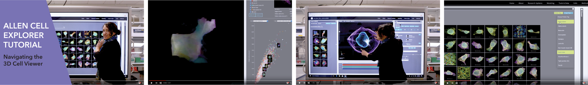
 We have released a new tutorial video. The 3D Cell Viewer is a tool in the Allen Cell Explorer for viewing thousands of 3D images of cellular structures and organelles. Megan Riel-Mehan represents the Animated Cell group to present an overview of several major upgrades and an easier to use interface for the 3D Cell Viewer tool.  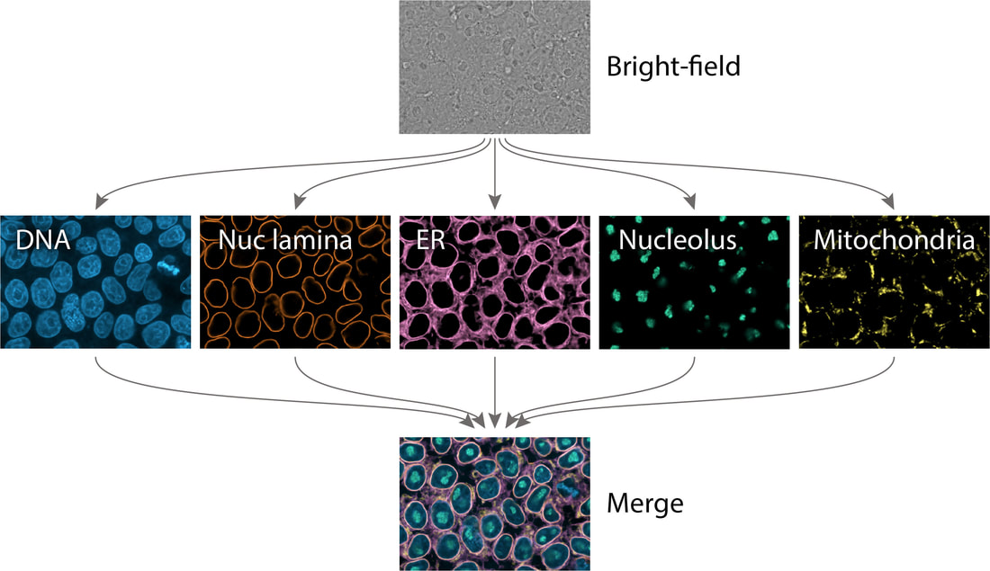 The Allen Institute for Cell Science has developed a tool for the prediction of fluorescently labeled structures in live cells solely from 3D brightfield microscopy images. This approach can be used to predict several structures of interest from the same 3D brightfield image, and can potentially be used in applications as diverse as cross-modal image registration, quantification of live cell imaging, and determination of cell state changes. This approach avoids use of fluorescence microscopy, typically used to identify subcellular structures, and the accompanying dyes and proteins are often expensive, time-consuming, and damaging to the cells. Learn more about this project in our paper Three dimensional cross-modal image inference: label-free methods for subcellular structure prediction.
 In addition to hosting booth #639 in the exhibitor hall, researchers from the Allen Institute for Cell Science will present in several venues including:
We look forward to seeing you at the 2017 ASCB | EMBO Meeting in Philadelphia December 2–6! |
OverviewTo receive the Allen Institute e-newsletter, sign up here. Archives
April 2024
Categories |
 RSS Feed
The Institute |
Legal |
Help & contact |
Follow Us
|
Copyright © 2024 Allen Institute. All Rights Reserved.
|
|
See more on alleninstitute.org
|