ALLEN CELL EXPLORER
  • About
      Institute
      1. Our science: CellScapes
      2. Past foundational projects
      3. News feed
      4. About us
      5. Careers
  • Allen Cell Collection
      Order cells & plasmids
      1. Cell Catalog
      2. Disease Collection Cell Catalog
      3. Cell Catalog quickview
      4. Cell video shorts
      Lab methods
      1. Video protocols
      2. Written protocols
      3. Our methodology
      4. Support forum
      About our hiPS cells
      1. hiPS Cell Structure Overview
      2. Visual Guide to Human Cells
      3. Cell structure observations
      4. Why endogenous tagging?
      5. Differentiation into cardiomyocytes
      6. Genomics
      7. Download cell data: Images, genomics, & features
  • Data & Digital Tools
      General
      1. Tools & resources overview
      2. Download cell data (images, genomics, features)
      3. Code repositories & software
      Desktop tools
      1. Allen Cell & Structure Segmenter
      2. AGAVE 3D pathtrace image viewer
      Web tools
      1. BioFile Finder
      2. Cell Feature Explorer
      3. Integrated Mitotic Stem Cell
      4. └ Z-stack viewer
      5. └ 3D viewer
      Web tools (con't)
      1. Simularium viewer
      2. Timelapse Feature Explorer
      3. Visual Guide to Human Cells
      4. Vol-E (Web Volume Viewer)
      5. 3D Cell Viewer
  • Analysis & Modeling
      Allen Integrated Cell models
      1. Visual Guide to Human Cells
      2. Integrated Mitotic Stem Cell
      3. └ Z-stack viewer
      4. └ 3D viewer
      5. Allen Integrated Cell
      6. └ 3D Probabilistic Modeling
      7. └ Label-free Determination
      4D biology models
      1. Simularium viewer
      Methodologies
      1. Drug perturbation pilot study
      2. hiPS cells during mitosis
      3. Differentiation into cardiomyocytes
  • Publications
      Articles
      1. Publications
      2. Preprints
      Presentations
      1. Talks & posters
  • Education
      Educational resources
      1. All resources
      2. Teaching materials
      Online tools popular with teachers
      1. Visual Guide to Human Cells
      2. Integrated Mitotic Stem Cell
      3. 3D Cell Feature Explorer
      4. 3D Cell Viewer
      5. hiPS cell structure overview
  • Support
      Questions
      1. FAQs
      2. Forum
      Tutorials for digital tools
      1. Video tutorials
      2. Visual Guide tutorial
      3. AGAVE documentation
      Lab methods
      1. Video protocols
      2. Written protocols
      3. Our methodology
  • 🔍
      SEARCHBAR

Cell structure observations

Observations about microscopy videos for 16 of the cell lines available in our Cell Catalog & 3D Cell Viewer.

Actin structures visualized via ß-actin, α-actinin, & actinomyosin

3/21/2017

 
ß-actin
α-actinin
Myosin IIB
Figure. Movies of ß-actin, α-actinin & actinomyosin in various actin structures. Top row movies: Z-stacks of live hiPS cells expressing mEGFP-tagged ß-actin (left), α-actinin (center) and non-muscle myosin heavy chain IIB (right) imaged on a spinning-disk confocal microscope. Images start from the bottom of the cells and end at the top. 
Picture
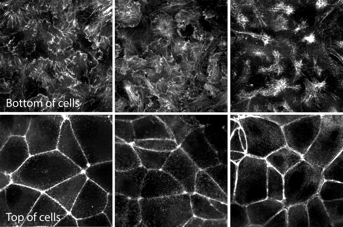
Figure 2. Images of the bottom and top of each structure for easier comparison. Representative images of mEGFP tagged ß-actin (left), α-actinin (center) and non-muscle myosin heavy chain IIB (right) from the bottom (top row) and tops (bottom row) of cells. These images are single slices taken from the z-stacks above. 
Observations
Actin - ß-actin
  • ß-actin comprises cytoskeletal filaments that drive protrusion and provide structural support and generate cellular force.
  • At the bottom of the cells, ß-actin is both in prominent filaments often associated with cell substrate adhesions and at the periphery of cell protrusions (lamellipodia).
  • Near the very top of the cells, ß-actin forms an apical actin band at cell-cell contacts, a feature common in epithelial cells. Small actin puncta of actin are visible along the top surface of cells.
  • ß-actin is also distributed diffusely throughout the cytoplasm and in brighter foci near cell cell contacts in the center of cells.

Actin bundles - α-actinin
  • α-actinin crosslinks actin filaments, forming actin bundles.
  • α-actinin localization is almost identical to ß-actin. The main difference is that α-actinin localizes less strongly to the lamellipodia at the protruding edges at the bottom of the cells, where there are fewer bundles.
  • Like ß-actin, α-actinin is particularly prominent in the actin-containing band near the top of the cell and in the filamentous structures at the bottom of the cell, along the substratum. 

Actomyosin bundles - Myosin IIB
  • Myosin IIB is a contractile motor protein that binds and crosslinks actin filaments.
  • Myosin IIB localization is similar to that of actin and α-actinin, residing in prominent filaments primarily at the bottom and top of cells.
  • At the bottom of cells it is primarily localized to the contractile stress fibers but not in the protruding regions of the cells.
  • Myosin IIB also localizes to the apical actin band found in epithelial cells. However, it is less obviously localized to the actin structures in the center of cells and does not form small puncta at the top surface.

In summary, α-actinin localizes to a subset of actin (e.g. the actin bundles) and myosin IIB to a yet smaller subset (actomyosin bundles) as expected. The general localization pattern of actin containing structures is consistent with an apical-basal epithelial polarity in these cells.

    About

    Observations and descriptions from the microscope

    Archives

    February 2019
    August 2018
    April 2018
    October 2017
    April 2017
    March 2017

    Categories

    All
    3-channel
    3D Rotation
    Actin
    Actinomyosin
    Actin Structures
    Adhesions
    Cardiomyocyte
    Cell Cell Contacts
    Cell-cell Contacts
    Cell Contacts
    Cell Division
    Cell Membrane
    Centrin
    Centrioles
    Chromatin
    Colony
    Connexin
    Connexin-43
    Desmoplakin
    Desmosomes
    DNA
    Endoplasmic Reticulum
    Endosome
    ER
    Fibrillarin
    FUS
    Gap Junctions
    Golgi Apparatus
    H2B
    Histone
    K-Ras
    Lamin
    Lamin B1
    LAMP1
    Lysosomes
    MEGFP
    Membrane
    Microtubule
    Mitochondria
    Mitosis
    MLC-2a
    MLC 2v
    MTagRFP-T
    Muscle
    Myosin
    Nuclear
    Nuclear Envelope
    Nuclear Pores
    Nucleolus
    Nucleophosmin
    Nucleoporin
    Nucleus
    Nup153
    Paraspeckles
    Paxillin
    Peroxisome
    Plasma Membrane
    PMP34
    Rab-5A
    Ras
    RNA-binding Protein
    Sarcomere
    Sarcomere M-line
    Sarcomere Thick Filaments
    Sarcoplasmic Reticulum
    Sec61
    SERCA2
    Sialyltransferase 1
    SMC 1A
    ß-actin
    ST6GAL1
    Stress Granules
    Tight Junctions
    Time Lapse
    Time Series
    Titin
    Tom20
    Troponin
    Tubulin
    ZO1
    Z Stack

    RSS Feed

The Institute

Home
Careers
Culture & Community
Archived Content

Legal

Terms of Use
Citation Policy
Privacy Policy
Cookie Settings

Help & contact

​FAQs
Help
​Send us a Message
​Sign up for our Newsletter
Allen Institute for Cell Science is a part of the Allen Institute. The mission of the Allen Institute is to understand the principles that govern life, and to advance health. Our creative and multi-dimensional teams focus on answering some of the biggest questions in bioscience. We accelerate foundational research, catalyze bold ideas, develop tools and models, and openly share our science to make a broad, transformational impact on the world.
Follow Us  
​Copyright © 2025 Allen Institute. All Rights Reserved.
  • About
      Institute
      1. Our science: CellScapes
      2. Past foundational projects
      3. News feed
      4. About us
      5. Careers
  • Allen Cell Collection
      Order cells & plasmids
      1. Cell Catalog
      2. Disease Collection Cell Catalog
      3. Cell Catalog quickview
      4. Cell video shorts
      Lab methods
      1. Video protocols
      2. Written protocols
      3. Our methodology
      4. Support forum
      About our hiPS cells
      1. hiPS Cell Structure Overview
      2. Visual Guide to Human Cells
      3. Cell structure observations
      4. Why endogenous tagging?
      5. Differentiation into cardiomyocytes
      6. Genomics
      7. Download cell data: Images, genomics, & features
  • Data & Digital Tools
      General
      1. Tools & resources overview
      2. Download cell data (images, genomics, features)
      3. Code repositories & software
      Desktop tools
      1. Allen Cell & Structure Segmenter
      2. AGAVE 3D pathtrace image viewer
      Web tools
      1. BioFile Finder
      2. Cell Feature Explorer
      3. Integrated Mitotic Stem Cell
      4. └ Z-stack viewer
      5. └ 3D viewer
      Web tools (con't)
      1. Simularium viewer
      2. Timelapse Feature Explorer
      3. Visual Guide to Human Cells
      4. Vol-E (Web Volume Viewer)
      5. 3D Cell Viewer
  • Analysis & Modeling
      Allen Integrated Cell models
      1. Visual Guide to Human Cells
      2. Integrated Mitotic Stem Cell
      3. └ Z-stack viewer
      4. └ 3D viewer
      5. Allen Integrated Cell
      6. └ 3D Probabilistic Modeling
      7. └ Label-free Determination
      4D biology models
      1. Simularium viewer
      Methodologies
      1. Drug perturbation pilot study
      2. hiPS cells during mitosis
      3. Differentiation into cardiomyocytes
  • Publications
      Articles
      1. Publications
      2. Preprints
      Presentations
      1. Talks & posters
  • Education
      Educational resources
      1. All resources
      2. Teaching materials
      Online tools popular with teachers
      1. Visual Guide to Human Cells
      2. Integrated Mitotic Stem Cell
      3. 3D Cell Feature Explorer
      4. 3D Cell Viewer
      5. hiPS cell structure overview
  • Support
      Questions
      1. FAQs
      2. Forum
      Tutorials for digital tools
      1. Video tutorials
      2. Visual Guide tutorial
      3. AGAVE documentation
      Lab methods
      1. Video protocols
      2. Written protocols
      3. Our methodology
  • 🔍
      SEARCHBAR