ALLEN CELL EXPLORER
  • About
      Institute
      1. News feed
      2. What we do
      3. Publications
      4. Allen Institute | allenInstitute.org
      5. Careers
      Site
      1. Home page
      2. Site updates
      3. Archived content
  • Allen Cell Collection
      Order cells & plasmids
      1. Cell Catalog
      2. Cell Catalog quickview
      3. Cell Shorts (documentaries on labs using our cells)
      4. Support forum
      Lab methods
      1. Instructional videos for success in the lab
      2. Standard operating procedures (written methods)
      3. Illustrated overviews
      About our hiPS cells
      1. hiPS Cell Structure Overview
      2. Visual Guide to Human Cells
      3. Cell structure observations
      4. Why endogenous tagging?
      5. Differentiation into cardiomyocytes
      6. Genomics
      7. Download cell data (images, genomics, features)
  • Data & Digital Tools
      Online image analysis
      1. Cell Feature Explorer (plotting & 3D viewer)
      2. 3D cell viewer (pre2018)
      3. Deep cell zoom (216,016 cells)
      Online modeling viewers
      1. Visual Guide to Human Cells
      2. Simularium (4D visual analysis)
      3. Integrated Mitotic Stem Cell
      4. └ Z-stack viewer
      5. └ 3D viewer
      6. Allen Integrated Cell viewer
      7. Label-free examples viewer
      8. 3D probabilistic model viewer
      Desktop tools
      1. Allen Cell & Structure Segmenter
      2. AGAVE 3D pathtrace image viewer
      Data & code
      1. Download cell data (images, genomics, features)
      2. Code repositories & software
  • Analysis & Modeling
      Allen Integrated Cell models
      1. Overview
      2. Integrated Mitotic Stem Cell
      3. └ Z-stack viewer
      4. └ 3D viewer
      5. Label-free Determination
      6. └ 3D viewer
      7. 3D Probabilistic Modeling
      8. └ 3D viewer
      9. Visual Guide to Human Cells
      4D biology models
      1. Simularium (online 4D viewer)
      Methodologies
      1. Drug perturbation pilot study
      2. hiPS cells during mitosis
      3. Differentiation into cardiomyocytes
  • Publications
      Articles
      1. All journal publications
      2. Preprints (biorxiv, arxiv)
      Posters
      1. Select posters
  • Education
      Education resources
      1. All Resources
      2. Teaching materials
      Online tools popular with teachers
      1. Visual Guide to Human Cells
      2. Integrated Mitotic Stem Cell
      3. Cell Feature Explorer (interactive plotting & 3D viewer)
      4. 3D cell viewer (pre2018 data)
      5. hiPS cell structure overview
  • Support
      Questions
      1. FAQs
      2. Forum
      Tutorials for digital tools
      1. Digital tool tutorials with videos
      2. Visual Guide tutorial
      3. AGAVE user guide
      Lab methods
      1. Instructional videos for success in the lab
      2. Standard operating procedures (written methods)
      3. Illustrated overviews
  • 🔍
      SEARCHBAR

Cell Structure Observations

Observations about microscopy videos for each of the 16 cell lines available in our Cell Catalog & 3D Cell Viewer.

Histones visualized via Histone H2B type 1-J

8/1/2018

 
Movie. Z-stack of live hiPS cell colony expressing mEGFP-tagged Histone H2B type 1-J protein. Cells were imaged in 3D on a spinning-disk confocal microscope. Movie starts at the bottom of the cells and ends at the top. Scale bar, 5µm.
Movie. Time-lapse movie of live hiPS cell colony expressing mEGFP-tagged Histone H2B type 1-J protein. Cells were imaged in 3D on a spinning-disk confocal microscope every 3 min. A single mid-level z-section is shown. Movie plays at 1260x real time. Scale bar, 5 µm.
Movie. ​Z-stack of live hiPS cell colony expressing mEGFP-tagged Histone H2B type 1-J protein. Cell Mask Deep Red Plasma Membrane dye was applied to mark cell boundaries (magenta). NucBlue Live Ready Probe Reagent was used to compare mEGFP-tagged H2B localization (left panel) to a NucBlue Live (Hoechst) DNA stain (left panel). Cells were imaged in 3D on a spinning-disk confocal microscope. Movie starts at the bottom of the cells and ends at the top. Scale bar, 5µm.
Observations
  • Histone H2B type 1-J is a protein that participates in organizing DNA. It and other histone proteins form a core around which DNA wraps. These structures, called nucleosomes, are the fundamental repeating units within chromatin (DNA and histones). The packing of these structures impacts DNA accessibility, affecting functions like transcription, replication, and repair of DNA.
  • In hiPS cells imaged with spinning-disk light microscopy, mEGFP-tagged Histone H2B type 1-J appears within the nucleus as puncta of varying size and intensity, likely reflecting more densely packed regions of nucleosomes, and in a “haze”, where nucleosomes cannot be resolved.
  • The localization of mEGFP-tagged Histone H2B type 1-J is similar to the pattern of the DNA-binding dye Hoechst. Hoechst binds the minor grove of DNA (with preference for A-T rich regions), while mEGFP-tagged Histone H2B type 1-J localizes to the nucleosomal cores around which the DNA wraps.
  • Histone H2B type 1-J pattern of localization and intensity changes through the cell cycle. During interphase, Histone H2B type 1-J localizes throughout the nucleus, with some regions of greater intensity (reflecting a greater density of protein) near the nuclear periphery and the nucleolus. During cell division, Histone H2B type 1-J localizes to chromosomes.

Nuclear pores visualized via Nucleoporin Nup153

8/1/2018

 
Movie. Z-stack of live hiPS cell colony expressing mEGFP-tagged Nucleoporin Nup153 protein. Cells were imaged in 3D on a spinning-disk confocal microscope. Movie starts at the bottom of the cells and ends at the top. Scale bar, 5µm.
Movie. Time-lapse movie of live hiPS cell colony expressing mEGFP-tagged Nucleoporin Nup153 protein. Cells were imaged in a 3D volume spanning ~1.5 µm through the middle of the cells on a spinning-disk confocal microscope every 3 min. A single mid-level z-section is shown. Movie plays at 1800x real time. Scale bar, 5 µm.
Observations
  • Nucleoporin Nup153 is a protein within the nuclear pore complex (NPC). The NPC bridges the double membrane of the nuclear envelope to regulate the exchange of molecules between the nucleus and cytoplasm. Nucleoporin Nup153 is located on the nuclear side of the NPC. It is critically involved in the ability of the NPC to transport molecules between the nucleus and cytoplasm; but it also plays other roles in nuclear properties and functions, including the shape of the NPC and the nucleus, and regulation of transcription.
  • In hiPS cells, Nucleoporin Nup153 appears as numerous dense puncta along the nuclear envelope, including within invaginations.
  • During cell division, Nucleoporin Nup153 localization changes with the reorganization of the nuclear envelope. During mitosis, Nuceloporin Nup153 localizes in a dim wavy structure, likely the ER. At the end of mitosis, Nucleoporin Nup153 appears on chromosomes before finally reappearing at the nuclear envelope.

Nucleolus visualized via nucleophosmin

4/24/2018

 
Movie. Z-stack and timelapse movie of nucleoli. The left panel shows a Z-stack of a live hiPS cell colony expressing mEGFP-tagged nucleophosmin. Cells were imaged in 3D on a spinning-disk confocal microscope. Right panel is the same image as the left but with contrast enhanced to visualize dimmer localization in mitotic cells. Movie starts at the bottom of the cells and ends at the top. Scale bar = 5µm.

Movie. Z-stack and timelapse movie of nucleoli. Timelapse movie of a live hiPS cell colony expressing mEGFP-tagged nucleophosmin. Cells were imaged on a spinning-disk confocal microscope every 3 min. Image is a maximum intensity projection through the volume of the cells. Frames were histogram matched to adjust for photobleaching. Movie plays at 900x real time. Scale bar = 5 µm.

Observations
  • Nucleophosmin marks the granular component of the nucleolus, the nuclear subcompartment where ribosomes are made.
  • During much of interphase the nucleolus exists in one to several large, textured clusters within the nucleus of hiPS cells. During cell division, the nucleolus appears to ‘melt’ and then dissociate. After cell division, the nucleolus reassembles, first into small particles and progressing into the larger textured clusters observed during interphase.
  • The mEGFP-tagged nucleophosmin exhibits many similarities with the mEGF-tagged fibrillarin, another marker of the nucleolus. However, it also exhibits notable differences, including its specific localization within the nucleolus, as each line marks a different component of the nucleus. The granular component of the nucleolus where nucleophosmin resides forms hollow-looking spheres, giving the nucleolus a mesh-like appearance when imaged with this marker.​

Nucleolus via Fibrillarin 

3/16/2017

 
Z-stack
​High magnification timelapse (cell division)
Figure 1. Movies of fibrillarin in Nucleoli. Left: Z-stack of live hiPS cells expressing mEGFP tagged fibrillarin imaged on a spinning-disk confocal microscope. Images start from the bottom of the cells and end at the top. Right: Timelapse movie of live hiPS cells expressing mEGFP tagged fibrillarin. Images were collected in 3D every 3 minutes for 1.5 hours on a spinning-disk confocal microscope. Image is a maximum intensity projection. Playback speed is 900x real time.
Picture
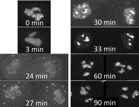
Figure 2. Time series of cell division. A single cell going through cell division taken from the movie on the right.
Observations
  • Fibrillarin marks the dense fibrillar component (DFC) of the nucleolus, the nuclear subcompartment where ribosome biogenesis occurs.
  • During much of interphase the nucleolus exists in 1-2 large, textured clusters within the nucleus of hiPS cells. During cell division, the nucleolus appears to ‘melt’ and then dissociate. After cell division, the nucleolus reassembles, first into small particles and progressing into the larger textured clusters observed during interphase. Low levels of fibrillarin are visible on chromosomes during chromosome segregation in mitosis.

Nuclear envelope visualized via lamin B1

3/10/2017

 
​Low magnification timelapse
​High magnification timelapse
Figure. Timelapse movies of hiPSC cells expressing mEGFP tagged Lamin B1. Images were collected in 3D every 3 minutes for 12 hours (left) or every 35 seconds for 23 minutes (right) on a spinning-disk confocal microscope. Images are maximum intensity projection (left) or single slices from the middle of the z-stack (right). Playback speed is 1800x (left) and 350x (right) real time.

Observations
  • Lamin B1 is a member of the lamin family of proteins that make up the nuclear lamina, located just inside the inner nuclear envelope. - In hiPS cells, nuclei in these cells occupy 30-50% of the cell volume making them very prominent. In the center of cells the nuclei occupy almost the entire cytoplasm such that a ‘bird’s eye view’ of the cell monolayer shows nuclei that appear tightly packed together.
  • As the cells enter mitosis, the lamin B1-containing nuclear envelope is seen to ruffle and take on a wavy morphology as it begins to breakdown. However the nuclear envelope does not completely breakdown during mitosis. As the nuclear envelope reforms, invaginations are seen that can look like spots within the nucleus. These spots decrease and disappear over time suggesting that they may be cell cycle related. These invaginations are also seen with Sec61B, which labels the ER including the peripheral ER surrounding the nucleus (See ER section).

    About

    Observations and descriptions from the microscope

    Archives

    February 2019
    August 2018
    April 2018
    October 2017
    April 2017
    March 2017

    Categories

    All
    3-channel
    3D Rotation
    Actin
    Actinomyosin
    Actin Structures
    Adhesions
    Cardiomyocyte
    Cell Cell Contacts
    Cell-cell Contacts
    Cell Contacts
    Cell Division
    Cell Membrane
    Centrin
    Centrioles
    Chromatin
    Colony
    Connexin
    Connexin-43
    Desmoplakin
    Desmosomes
    DNA
    Endoplasmic Reticulum
    Endosome
    ER
    Fibrillarin
    FUS
    Gap Junctions
    Golgi Apparatus
    H2B
    Histone
    K-Ras
    Lamin
    Lamin B1
    LAMP1
    Lysosomes
    MEGFP
    Membrane
    Microtubule
    Mitochondria
    Mitosis
    MLC-2a
    MLC 2v
    MTagRFP-T
    Muscle
    Myosin
    Nuclear
    Nuclear Envelope
    Nuclear Pores
    Nucleolus
    Nucleophosmin
    Nucleoporin
    Nucleus
    Nup153
    Paraspeckles
    Paxillin
    Peroxisome
    Plasma Membrane
    PMP34
    Rab-5A
    Ras
    RNA-binding Protein
    Sarcomere
    Sarcomere M-line
    Sarcomere Thick Filaments
    Sarcoplasmic Reticulum
    Sec61
    SERCA2
    Sialyltransferase 1
    SMC 1A
    ß-actin
    ST6GAL1
    Stress Granules
    Tight Junctions
    Time Lapse
    Time Series
    Titin
    Tom20
    Troponin
    Tubulin
    ZO1
    Z Stack

    RSS Feed

Home

Terms of Use

Citation Policy

Privacy Policy

FAQs

Help

Send us a message

Our diversity, equity & inclusion commitment
Copyright © 2023 Allen Institute. All Rights Reserved.
cellscience.alleninstitute.org
  • About
      Institute
      1. News feed
      2. What we do
      3. Publications
      4. Allen Institute | allenInstitute.org
      5. Careers
      Site
      1. Home page
      2. Site updates
      3. Archived content
  • Allen Cell Collection
      Order cells & plasmids
      1. Cell Catalog
      2. Cell Catalog quickview
      3. Cell Shorts (documentaries on labs using our cells)
      4. Support forum
      Lab methods
      1. Instructional videos for success in the lab
      2. Standard operating procedures (written methods)
      3. Illustrated overviews
      About our hiPS cells
      1. hiPS Cell Structure Overview
      2. Visual Guide to Human Cells
      3. Cell structure observations
      4. Why endogenous tagging?
      5. Differentiation into cardiomyocytes
      6. Genomics
      7. Download cell data (images, genomics, features)
  • Data & Digital Tools
      Online image analysis
      1. Cell Feature Explorer (plotting & 3D viewer)
      2. 3D cell viewer (pre2018)
      3. Deep cell zoom (216,016 cells)
      Online modeling viewers
      1. Visual Guide to Human Cells
      2. Simularium (4D visual analysis)
      3. Integrated Mitotic Stem Cell
      4. └ Z-stack viewer
      5. └ 3D viewer
      6. Allen Integrated Cell viewer
      7. Label-free examples viewer
      8. 3D probabilistic model viewer
      Desktop tools
      1. Allen Cell & Structure Segmenter
      2. AGAVE 3D pathtrace image viewer
      Data & code
      1. Download cell data (images, genomics, features)
      2. Code repositories & software
  • Analysis & Modeling
      Allen Integrated Cell models
      1. Overview
      2. Integrated Mitotic Stem Cell
      3. └ Z-stack viewer
      4. └ 3D viewer
      5. Label-free Determination
      6. └ 3D viewer
      7. 3D Probabilistic Modeling
      8. └ 3D viewer
      9. Visual Guide to Human Cells
      4D biology models
      1. Simularium (online 4D viewer)
      Methodologies
      1. Drug perturbation pilot study
      2. hiPS cells during mitosis
      3. Differentiation into cardiomyocytes
  • Publications
      Articles
      1. All journal publications
      2. Preprints (biorxiv, arxiv)
      Posters
      1. Select posters
  • Education
      Education resources
      1. All Resources
      2. Teaching materials
      Online tools popular with teachers
      1. Visual Guide to Human Cells
      2. Integrated Mitotic Stem Cell
      3. Cell Feature Explorer (interactive plotting & 3D viewer)
      4. 3D cell viewer (pre2018 data)
      5. hiPS cell structure overview
  • Support
      Questions
      1. FAQs
      2. Forum
      Tutorials for digital tools
      1. Digital tool tutorials with videos
      2. Visual Guide tutorial
      3. AGAVE user guide
      Lab methods
      1. Instructional videos for success in the lab
      2. Standard operating procedures (written methods)
      3. Illustrated overviews
  • 🔍
      SEARCHBAR