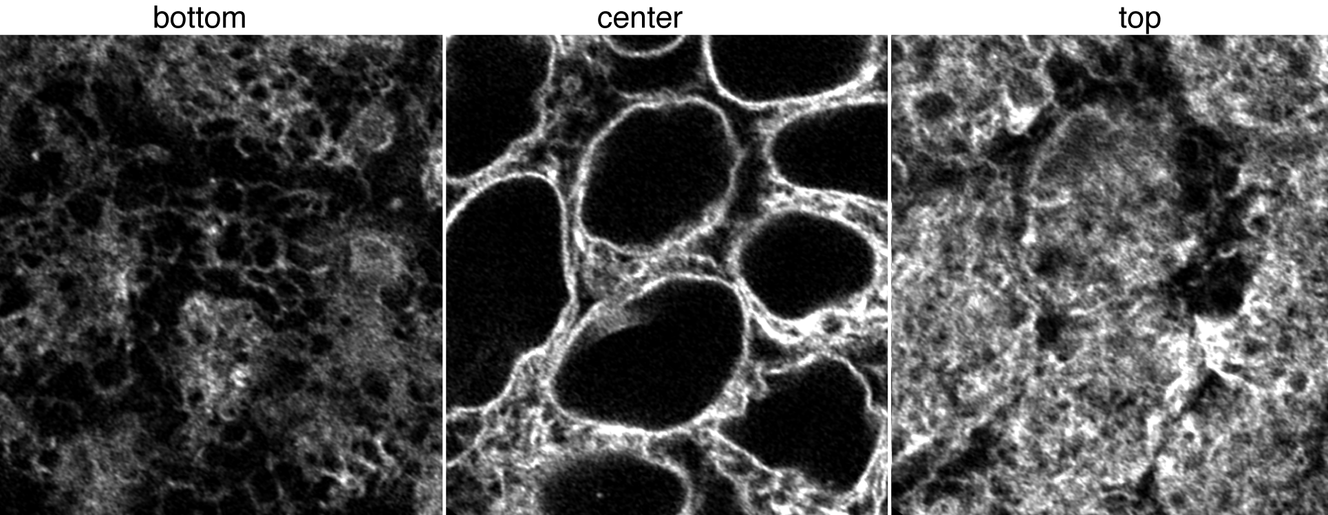
Figure 1. Movies of Sec61-ß in ER. Top left: Z-stack of live hiPS cells expressing mEGFP tagged Sec61-ß imaged on a spinning-disk confocal microscope. Images start from the bottom of the cells and end at the top. Top right: Timelapse movie of live hiPS cells expressing mEGFP tagged Sec61-ß. Images were collected in 3D every 2 minutes for 3 hours on a spinning-disk confocal microscope. Image is a single slice through the center of the cells. Playback speed is 600x real time. Bottom image panel: live hiPSC cells expressing mEGFP tagged Sec61-ß imaged on a Zeiss LSM 880 AiryScan FAST in super-resolution mode. Figure 2. Images of Sec61-ß in ER. Left, middle, and right images represent a single slice at the bottom, center, and top of cells with AiryScanFast SuperRes Observations
Comments are closed.
|
AboutObservations and descriptions from the microscope Archives
February 2019
Categories
All
|
The Institute |
Legal |
Help & contact |
Follow Us
|
Copyright © 2024 Allen Institute. All Rights Reserved.
|
|
See more on alleninstitute.org
|


 RSS Feed